Pupil
2008/9 Schools Wikipedia Selection. Related subjects: Health and medicine

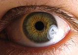
The pupil is the central transparent area (showing as black). The greenish-brown area surrounding it is the iris. The white outer area is the sclera, the central transparent part of which is the cornea.
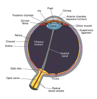
The pupil is the opening that is located in the centre of the iris of the eye and that controls the amount of light that enters the eye. It appears black because most of the light entering the pupil is absorbed by the tissues inside the eye. In optical terms, the anatomical pupil is the eye's aperture and the iris is the aperture stop. The image of the pupil as seen from outside the eye is the entrance pupil, which does not exactly correspond to the location and size of the physical pupil because it is magnified by the cornea.
Comparative anatomy
In humans and many animals (but few fish), the size of the pupil is controlled by involuntary constriction and dilation of the iris in order to regulate the intensity of light entering the eye. This is known as the pupillary reflex. In normal room light, a healthy human pupil has a diameter of about 3–4 millimeters, in bright light, the pupil has a diameter of about 1.5 millimeters, and in dim light the diameter is enlarged to about 8 millimeters. The narrowing of the pupil results in a greater focal range. (see aperture for a more detailed explanation)
The shape of the pupil varies between species. Common shapes are circular or slit-shaped, although more convoluted shapes can be found in aquatic species. The reasons for the variation in shapes are complex; the shape is closely related to the optical characteristics of the lens, the shape and sensitivity of the retina, and the visual requirements of the species.
Slit-shaped pupils are found in species which are active in a wide range of light levels. In strong light, the pupil constricts and is small, but still allows light to be cast over a large part of the retina.
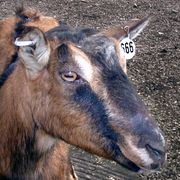
The orientation of the slit may be related to the direction of motions the eye is required to notice most sensitively (so a vertical pupil would increase the sensitivity of the eyes of a small cat to the horizontal scurrying of mice). The narrower the pupil, the more accurate the depth perception of peripheral vision is, so narrowing it in one direction would increase depth perception in that plane. Animals like goats and sheep may have evolved horizontal pupils because better vision in the vertical plane may be beneficial in mountainous environments.
Many snakes, such as boas, pythons and vipers, have vertical, slit-shaped pupils that help them to hunt prey under a wide range of light conditions. Small cats and foxes also have slit shaped pupils while lions and wolves have round pupils even though they are in the same respective families. Some hypothesize that this is because slit pupils are more beneficial for animals that hunt small prey rather than large prey.
When an eye is photographed with a flash, the iris cannot close the pupil fast enough and the blood-rich retina is illuminated, resulting in the red-eye effect.
Constriction of the pupil
When bright light is shone on the eye, it will automatically constrict. This is the pupillary reflex, which is an important test of brainstem function. Furthermore, the pupil will dilate if a person sees an object of interest.
The oculomotor nerve, specifically the parasympathetic part coming from the Edinger-Westphal nucleus, terminates on the circular iris sphincter muscle. When this muscle contracts, it reduces the size of the pupil.
The iris is a contractile structure, consisting mainly of smooth muscle, surrounding the pupil. Light enters the eye through the pupil, and the iris regulates the amount of light by controlling the size of the pupil. The iris contains two groups of smooth muscles; a circular group called the sphincter pupillae, and a radial group called the dilator pupillae. When the sphincter pupillae contract, the iris decreases or constricts the size of the pupil. The dilator pupillae, innervated by sympathetic nerves from the superior cervical ganglion, cause the iris to dilate when they contract. These muscles are sometimes referred to as intrinsic eye muscles.
Certain drugs cause constriction of the pupils, such as alcohol and opiates. Other drugs, such as atropine and amphetamines cause pupil dilation.
Another term for the constriction of the pupil is miosis. Substances that cause miosis are described as miotic.

