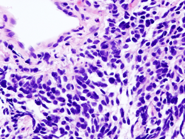
Image:Lung small cell carcinoma (1) by core needle biopsy.jpg
From Wikipedia, the free encyclopedia

No higher resolution available.
Lung_small_cell_carcinoma_(1)_by_core_needle_biopsy.jpg (600 × 452 pixels, file size: 343 KB, MIME type: image/jpeg)
File history
Click on a date/time to view the file as it appeared at that time.
| Date/Time | Dimensions | User | Comment | |
|---|---|---|---|---|
| current | 05:58, 15 January 2006 | 600×452 (343 KB) | KGH | (Histopathologic image of small cell carcinoma of the lung. CT-guided core needle biopsy. H & E stain. ==Licensing== {{GFDL-self}} category:histopathology) |
File links
The following pages on Schools Wikipedia link to this image (list may be incomplete):
Metadata
This file contains additional information, probably added from the digital camera or scanner used to create or digitize it. If the file has been modified from its original state, some details may not fully reflect the modified file.
| Camera manufacturer | OLYMPUS OPTICAL CO.,LTD. |
|---|---|
| Camera model | DP70 |
| Exposure time | 1/34 sec (0.029411764705882) |
| Date and time of data generation | 2005/03/30 16:32:10 |
| Image title | OLYMPUS DIGITAL CAMERA |
| Horizontal resolution | 72 dpi |
| Vertical resolution | 72 dpi |
| File change date and time | 2005/03/30 16:32:10 |
| Y and C positioning | 2 |
| ISO speed rating | 200 |
| Exif version | 2 |
| Date and time of digitizing | 2005/03/30 16:32:21 |
| Shutter speed | 5.587 |
| Metering mode | Spot |
| Light source | Unknown |
| Colour space | sRGB |
| Sensing method | One-chip colour area sensor |
