Image:Histology bse.jpg
From Wikipedia, the free encyclopedia
Size of this preview: 602 × 480 pixels
Full resolution (700 × 558 pixels, file size: 73 KB, MIME type: image/jpeg)
| |
This is a file from the Wikimedia Commons. The description on its description page there is shown below. |
 |
This image is a work of a United States Department of Agriculture employee, taken or made during the course of the person's official duties. As a work of the U.S. federal government, the image is in the public domain. |  |
| Description |
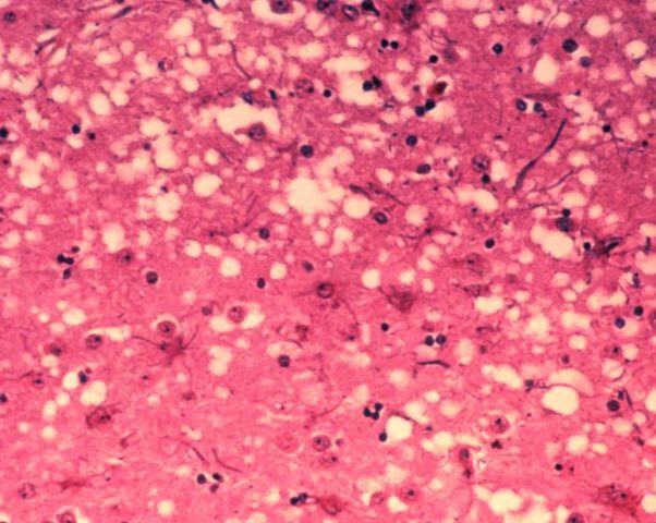
English: This micrograph of brain tissue reveals the cytoarchitectural histopathologic changes found in bovine spongiform encephalopathy. The presence of vacuoles, i.e. microscopic “holes” in the gray matter, gives the brain of BSE-affected cows a sponge-like appearance when tissue sections are examined in the lab.
Nederlands: Deze microscopische opname toont hersenweefsel van een koe die aan BSE gestorven is. Tussen de hersencellen ziet men duidelijk verschillende vacuoles, die deze coupe (weefselsnede) een sponsachtig uitzicht geven.
|
|---|---|
| Source |
Public Health Image Library, APHIS: http://www.aphis.usda.gov/lpa/issues/bse/bse_photogallery.html |
| Date |
2003 |
| Author |
Dr. Al Jenny |
| Permission ( Reusing this image) |
see below |
| Other versions | http://en.wikipedia.org/wiki/Image:Aphis.usda.gov_BSE_5.jpg |
File history
Click on a date/time to view the file as it appeared at that time.
| Date/Time | Thumbnail | Dimensions | User | Comment | |
|---|---|---|---|---|---|
| current | 10:21, 21 June 2005 |  |
700×558 (73 KB) | Obarskyr | ({{PD}} ) |
File links
The following pages on Schools Wikipedia link to this image (list may be incomplete):