Image:FluorescentCells.jpg
From Wikipedia, the free encyclopedia
Size of this preview: 480 × 480 pixels
Full resolution (512 × 512 pixels, file size: 56 KB, MIME type: image/jpeg)
| |
This is a file from the Wikimedia Commons. The description on its description page there is shown below. |
Contents |
Information
| Description |
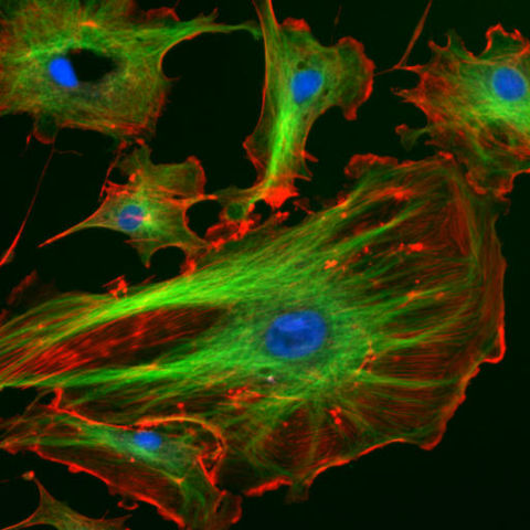
English: Endothelial cells under the microscope. Nuclei are stained blue with DAPI, microtubles are marked green by an antibody bound to FITC and actin filaments are labelled red with phalloidin bound to TRITC. Bovine pulmonary arthery endothelial cells
Deutsch: Endothelzellen unter dem Mikroskop. Die Zellkerne sind mit DAPI blau markiert. Die Mikrotubuli wurden über einen Antikörper grün markiert. Mit rot fluoreszierendem Phalloidin wurden die Aktinfilamente markiert.
Français : Cellulles endothéliales vues au microscope. En bleu, noyaux marqués au DAPI. En vert, microtubules marqués par un anticorps. En rouge, actine marquée à la phalloïdine.
Lietuvių: Citoskeletas. Aktino filamentai - raudina, mikrotubulės - žalia, branduolys - melyna.
Română: Sub microscop Celule endoteliale . microtubulii sunt de culoare verde, iar filamentele de actină sunt roşii, pe când nucleul celulei este colorat albastru
Русский: Цитоскелет эукариот. Актиновые микрофиламенты окрашены в красный, микротрубочки — в зеленый, ядра клеток — в голубой цвет.
Українська: Цитоскелет еукаріот. Актинові мікрофіламенти забарвлені в червоний колір, мікротрубочки — в зелений, ядра кліток — в блакитний
|
|---|---|
| Source |
http://rsb.info.nih.gov/ij/images/ |
| Date | |
| Author | |
| Permission ( Reusing this image) |
example image from the ImageJ-Programmpaket (public domain)
|
Original file
This image has been taken from the German Wikipedia
The original uploader is de:Benutzer:Jan R. The original upload was at 4th December 2005.
Original description
Endothelzellen unter dem Mikroskop. Die Zellkerne sind mit DAPI blau markiert. Die Mikrotubuli wurden über einen Antikörper grün markiert. Mit rot fluoreszierendem Phalloidin wurden die Aktinfilamente markiert.
Quelle: Beispielsbild aus dem ImageJ-Programmpaket (public domain), siehe http://rsb.info.nih.gov/ij/
Licensing
 |
This work is in the public domain in the United States because it is a work of the United States Federal Government under the terms of Title 17, Chapter 1, Section 105 of the US Code. See Copyright. Note: This only applies to works of the Federal Government and not to the work of any individual U.S. state, territory, commonwealth, county, municipality, or any other subdivision. This template also does not apply to postage stamps published by the United States Postal Service after 1978. (See 206.02(b) of Compendium II: Copyright Office Practices). العربية | Български | Česky | Deutsch | English | Español | Français | Magyar | Italiano | 日本語 | 한국어 | Polski | Português | 中文(繁體) | 中文(简体) | +/- |
 |
File history
Click on a date/time to view the file as it appeared at that time.
| Date/Time | Dimensions | User | Comment | |
|---|---|---|---|---|
| current | 15:07, 24 March 2006 | 512×512 (56 KB) | Splette | ({{Information |Description = Endothelial cells under the microscope. Nuclei are stained blue with DAPI, microtubles are marked green by an antibody and actin filaments are labelled red with phalloidin. |Source = http://rsb.info.nih.gov/ij |Date = |Author) |