Image:Epithelial-cells.jpg
From Wikipedia, the free encyclopedia
No higher resolution available.
Epithelial-cells.jpg (202 × 202 pixels, file size: 46 KB, MIME type: image/jpeg)
| |
This is a file from the Wikimedia Commons. The description on its description page there is shown below. |
-
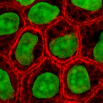
English: Cultured MDCK en:wikipedia:epithelial cells were stained for en:wikipedia:keratin, desmoplakin, and en:wikipedia:DNA. The stained cells were visualized by scanning laser confocal microscopy. The image shows how keratin cytoskeletal filaments are concentrated around the edge of the cells and merge into the desmoplakin which is located at en:wikipedia:desmosomes of the surface membrane. The network of keratin to desmosome to keratin linking the cells of an epithelial sheet is what holds together tissues like skin.
-
Slovenščina: Celice epitelija, obarvane za keratin, dezmoplakin in DNK.
- This image is taken from the wikibooks Cell Biology textbook (licensed under the GFDL): http://wikibooks.org/wiki/Image:Keratin.jpg
- The copyright to this image is retained by John Schmidt ( user:JWSchmidt).
- Permission is granted to copy, distribute and/or modify this image under the terms of the GFDL, as indicated in the fine print at the bottom of this page. If you do not want to use this image under the terms of the GFDL, you can alternatively use it under the terms of the Creative Commons Attribution-NonCommercial-ShareAlike License.
 |
Permission is granted to copy, distribute and/or modify this document under the terms of the GNU Free Documentation license, Version 1.2 or any later version published by the Free Software Foundation; with no Invariant Sections, no Front-Cover Texts, and no Back-Cover Texts. A copy of the license is included in the section entitled " GNU Free Documentation license". Aragonés | العربية | Asturianu | Беларуская (тарашкевіца) | Български | বাংলা | ইমার ঠার/বিষ্ণুপ্রিয়া মণিপুরী | Brezhoneg | Bosanski | Català | Cebuano | Česky | Dansk | Deutsch | Ελληνικά | English | Esperanto | Español | Eesti | Euskara | فارسی | Suomi | Français | Gaeilge | Galego | עברית | Hrvatski | Magyar | Bahasa Indonesia | Ido | Íslenska | Italiano | 日本語 | ქართული | ភាសាខ្មែរ | 한국어 | Kurdî / كوردی | Latina | Lëtzebuergesch | Lietuvių | Bahasa Melayu | Nnapulitano | Nederlands | Norsk (nynorsk) | Norsk (bokmål) | Occitan | Polski | Português | Română | Русский | Slovenčina | Slovenščina | Shqip | Српски / Srpski | Svenska | తెలుగు | ไทย | Tagalog | Türkçe | Українська | اردو | Tiếng Việt | Volapük | Yorùbá | 中文(简体) | 中文(繁體) | +/- |
This image can be used under the terms of either the GFDL or the Creative Commons Attribution-NonCommercial-ShareAlike License.
File history
Click on a date/time to view the file as it appeared at that time.
| Date/Time | Dimensions | User | Comment | |
|---|---|---|---|---|
| current | 19:49, 2 May 2005 | 202×202 (46 KB) | Helix84 | (Cultured MDCK epithelial cells were stained for keratin, desmoplakin, and DNA. The stained cells were visualized by scanning laser confocal microscopy. The image shows how keratin [[Cytoskeleton|cytoskele) |
File links
The following file is a duplicate of this file: