From Wikipedia, the free encyclopedia
 Size of this preview: 597 × 480 pixels
 Full resolution (1,560 × 1,254 pixels, file size: 518 KB, MIME type: image/jpeg)
 |
This is a file from the Wikimedia Commons. The description on its description page there is shown below.Commons is a freely licensed media file repository. You can help.
|
| Description |
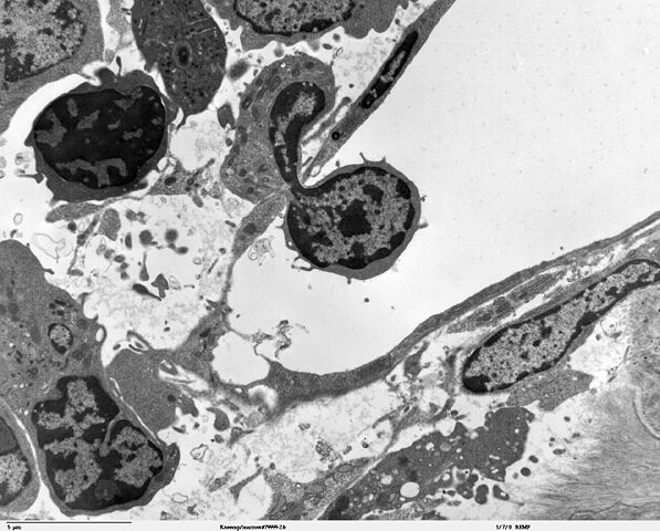
Transmission electron micrscope image of a thin section cut through an area of bone marrow area near the cartilage/bone interface in a mouse kneecap. Image shows small opening in the thin endotheliun of the vascular sinus wall, where a blood cell is crossing the thin vascular sinus wall and into the sinus lumen. JEOL 100CX TEM |
| Source |
|
| Date |
|
| Author |
Louisa Howard, Roy Fava |
Permission
( Reusing this image) |
PD
|
Licensing
 |
This file has been (or is hereby) released into the public domain by its author, Louisa Howard and Roy Fava. This applies worldwide. In case this is not legally possible:
Louisa Howard and Roy Fava grants anyone the right to use this work for any purpose, without any conditions, unless such conditions are required by law.
Afrikaans | Anglo-Saxon | العربية | Беларуская | Беларуская (тарашкевіца) | Български | Česky | Словѣ́ньскъ / ⰔⰎⰑⰂⰡⰐⰠⰔⰍⰟ | Dansk | Deutsch | ދިވެހިބަސް | ཇོང་ཁ | Ελληνικά | English | Español | فارسی | Suomi | Français | Avañe'ẽ | Հայերեն | Italiano | 日本語 | 한국어 | Plattdüütsch | Nederlands | Norsk (bokmål) | ਪੰਜਾਬੀ | Polski | Português | Română | Русский | Svenska | தமிழ் | Türkçe | Українська | Volapük | 中文 | 中文(台灣) | 粵語 | +/- |
File history
Click on a date/time to view the file as it appeared at that time.
|
|
Date/Time |
Dimensions |
User |
Comment |
| current |
14:25, 7 October 2006 |
1,560×1,254 (518 KB) |
Patho |
|
File links
The following pages on Schools Wikipedia link to this image (list may be incomplete):

