Image:Infiltrating ductal carcinoma of the breast.jpg

From Wikipedia, the free encyclopedia

Wikimedia Commons logo This is a file from the Wikimedia Commons. The description on its description page there is shown below.
Commons is a freely licensed media file repository. You can help.
Description

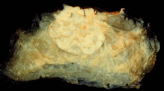
Infiltrating ductal carcinoma of the breast

This breast carcinoma, seen here in a mastectomy specimen, is unusually large by today's standards. The tumor measures 5 cm in greatest dimension and extensively involves the nipple (which can be seen as the papillated surface at the top of the specimen. The only way such a superficial tumor can get so large is for the patient to ignore it and not seek medical attention.

I have never had much luck in getting a good gross picture of a breast cancer; such photos are so dominated by shiny, greasy adipose tissue that the tumor gets lost in the fat. In this case I tried something different.

On receipt of the specimen, I fixed it overnight in formalin. Then, as part of the examination, I breadloafed the specimen along parasagittal planes. This section is the one that went through the nipple. Because the tumor was so big, I could retain this slice and not compromise the sampling for the microscopic exam. Then, I pinned this slice out in a wax pan and fixed it an additonal night in formalin. By then, the slice was as rigid as it was likely to get. I soaked the slice for three more nights in Carnoy's fluid to extract the fat. When there was no more observable fat, I dried the slice with a towel and photographed it. Certainly, the absence of fat gives the native breast tissue an unnatural appearance, but the anatomic relationship between the tumor and the surrounding breast is readily appreciated.

This photo was shot with a Minolta X-370 with bellows lens, on Ektachrome Elite 100 film, daylight balance, using a blue compensation filter. I believe the exposure was something like f/8 at 1/4 second.

Photograph by Ed Uthman, MD. Public domain. Posted 27 May 01

Source

http://web2.airmail.net/uthman/specimens/index.html

Date
Author
Permission
( Reusing this image)

PD


Licensing

Public domain This file has been (or is hereby) released into the public domain by its author, Ed Uthman. This applies worldwide.

In case this is not legally possible:
Ed Uthman grants anyone the right to use this work for any purpose, without any conditions, unless such conditions are required by law.


Afrikaans | Anglo-Saxon | العربية | Беларуская | Беларуская (тарашкевіца) | Български | Česky | Словѣ́ньскъ / ⰔⰎⰑⰂⰡⰐⰠⰔⰍⰟ | Dansk | Deutsch | ދިވެހިބަސް | ཇོང་ཁ | Ελληνικά | English | Español | فارسی | Suomi | Français | Avañe'ẽ | Հայերեն | Italiano | 日本語 | 한국어 | Plattdüütsch | Nederlands | ‪Norsk (bokmål)‬ | ਪੰਜਾਬੀ | Polski | Português | Română | Русский | Svenska | தமிழ் | Türkçe | Українська | Volapük | 中文 | ‪中文(台灣)‬ | 粵語 | +/-

File history

Click on a date/time to view the file as it appeared at that time.

Date/Time Dimensions User Comment
current 10:17, 5 June 2006 550×305 (26 KB) Patho ({{Information| |Description=Infiltrating ductal carcinoma of the breast This breast carcinoma, seen here in a mastectomy specimen, is unusually large by today's standards. The tumor measures 5 cm in greatest dimension and extensively involves the nipple )
The following pages on Schools Wikipedia link to this image (list may be incomplete):
The 2008 Wikipedia for Schools is sponsored by SOS Children , and consists of a hand selection from the English Wikipedia articles with only minor deletions (see www.wikipedia.org for details of authors and sources). See also our Disclaimer.