Image:MitochondrionCAM.jpg
From Wikipedia, the free encyclopedia
No higher resolution available.
MitochondrionCAM.jpg (618 × 409 pixels, file size: 37 KB, MIME type: image/jpeg)
Summary
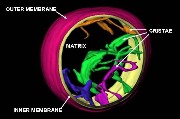
This is an image of a frozen-hydrated rat liver mitochondrion based on work done at the Wadsworth Centre's Resource for Visualization of Biological Complexity ( http://www.wadsworth.org/databank/electron/cryomito_dis2.html). It has not been previously published and I am releasing it into public domain for this article. The point is that the cristae are not folds of the inner membrane of the mitochondrion but pleomorphic invaginations with narrow tubular connections to each other and to the peripheral region of the inner membrane. I can advise on how to change your cartoon to be more accurate, while still retaining the basic format. There are numerous published references in last 10 years, the most recent: C.A. Mannella (2006) Biophysica et Biochimica Acta 1762: 140-147
Licensing
 |
This work has been released into the public domain by the copyright holder. This applies worldwide. In case this is not legally possible: |
File history
Click on a date/time to view the file as it appeared at that time.
| Date/Time | Dimensions | User | Comment | |
|---|---|---|---|---|
| current | 02:14, 17 July 2006 | 618×409 (37 KB) | Pschemp ( Talk | contribs) | (cropped version) |
| revert | 02:40, 5 February 2006 | 720×540 (28 KB) | Carmmann ( Talk | contribs) | (This is an image of a frozen-hydrated rat liver mitochondrion based on work done at the Wadsworth Centre's Resource for Visualization of Biological Complexity (http://www.wadsworth.org/databank/electron/cryomito_dis2.html). It has not been previously pub) |
See the setup instructions for more information.