From Wikipedia, the free encyclopedia
 Size of this preview: 368 × 480 pixels
 Full resolution (1,150 × 1,500 pixels, file size: 484 KB, MIME type: image/jpeg)
 |
This is a file from the Wikimedia Commons. The description on its description page there is shown below.Commons is a freely licensed media file repository. You can help.
|
| Description |
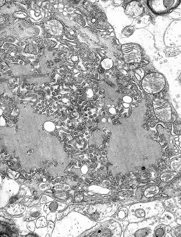
ID#: 1876 Description: Electron micrograph of the Rabies Virus. This electron micrograph shows the rabies virus, as well as Negri bodies, or cellular inclusions. Content Providers(s): CDC/Dr. Fred Murphy Creation Date: 1975 Copyright Restrictions: None - This image is in the public domain and thus free of any copyright restrictions. As a matter of courtesy we request that the content provider be credited and notified in any public or private usage of this image. |
| Source |
http://phil.cdc.gov/PHIL_Images/03082002/00012/PHIL_1876_lores.jpg high res.: |
| Date |
|
| Author |
This media is lacking author information.
العربية | Česky | Deutsch | English | Español | Suomi | Français | עברית | 日本語 | 한국어 | Lietuvių | Nederlands | Polski | Русский | 中文(简体) | 中文(繁體) | +/-
|
Permission
( Reusing this image) |
 |
This image is a work of the Centers for Disease Control and Prevention, part of the United States Department of Health and Human Services, taken or made during the course of an employee's official duties. As a work of the U.S. federal government, the image is in the public domain. |
|
|
File history
Click on a date/time to view the file as it appeared at that time.
|
|
Date/Time |
Dimensions |
User |
Comment |
| current |
17:56, 30 May 2006 |
1,150×1,500 (484 KB) |
Patho |
|
File links
The following pages on Schools Wikipedia link to this image (list may be incomplete):

